Health issues related to obesity can take many different forms. Obese people are more likely to have nonalcoholic steatohepatitis (NASH), a kind of fatty liver disease that can lead to cancer.
The lack of appropriate and effective techniques to separate and characterise hepatic stellate cells (HSCs), which have been shown to play a key role in the advancement of liver fibrosis and liver cancer in NASH patients, has contributed to the lack of treatment for NASH. With their novel method of performing cold enzymatic perfusion to separate HSCs from both murine and human fatty liver-associated malignancies, researchers from Osaka Metropolitan University made a significant advancement in their quest.
Cellular and Molecular Gastroenterology and Hepatology published their research.
Professor Naoko Ohtani, the study’s principal investigator, said that HSCs “promote obesity-associated liver cancer by producing senescence-associated secretory factors that decrease anti-tumour immunity in the tumour microenvironment.” The existing approach to isolating HSCs, however, is confined to normal livers, “hence, isolating and then characterising HSCs under varied situations is considered a precondition for explaining their roles in cancer progression.”
Because of the buildup of body fat, Professor Ohtani explained, “The present HSC isolation method conducts perfusion from the inferior vena cava, which is regrettably not apparent in fatty livers.” This technique is also carried out at a temperature of 37 °C, which may cause the expression of genes associated with heat stress and result in artefacts or inaccurate representations of tissue structures.
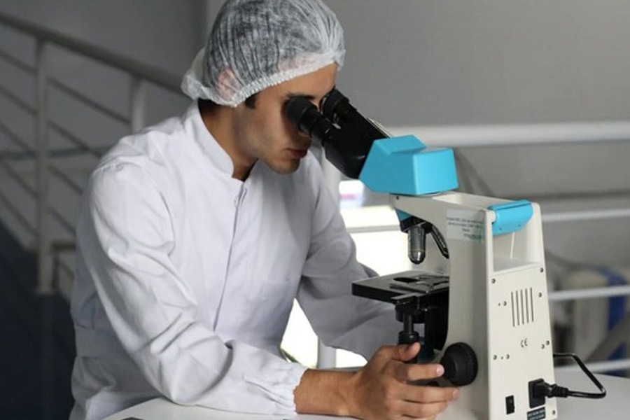
To address these issues, the research team made an effort to separate HSCs from the livers of mice given a normal diet and animals fed a high-fat diet that was cancerous. HSCs were gathered from both tumour and healthy tissue sites. The scientists used perfusion from the mice’s portal veins while using enzymatic solutions at a temperature of 6°C.
High-yield and high-purity HSCs were isolated by the researchers. Under varied conditions of either normal or fatty liver tissue, the portal vein was evident, and the cold enzymatic treatment reduced the production of artefacts. The experiment revealed CD49a to be a reliable HSC surface identifier, and an antibody against it made it possible to identify and gather HSCs with high CD49a expression.
The new technique also successfully isolates HSCs from liver cancer patients.
The researchers have published a number of studies on liver cancer linked to NASH, the most recent of which is this one. Professor Ohtani added, “After many tries and errors, we discovered this approach, which allowed highly effective extraction of high purity HSCs from fatty livers. “We anticipate that it will offer some resources for examining the function of HSCs and help in the development of therapies for NASH-associated liver cancer.”

കൈരളി ന്യൂസ് വാട്സ്ആപ്പ് ചാനല് ഫോളോ ചെയ്യാന് ഇവിടെ ക്ലിക്ക് ചെയ്യുക
Click Here